УЗИ мочевого пузыря
Модератор: KsV
-
А.М.Шифрин
- Сообщения: 60
- Зарегистрирован: 12 апр 2009, 18:41
- Контактная информация:
Re: УЗИ мочевого пузыря
Если не смещается, то действительно, полип. Но обязательна биопсия с гистологией.
Re: УЗИ мочевого пузыря
возможно, папиллярная опухоль мочеточника. необходим осмотр почки на гидронефроз. пионефроз - злок опухоль мочеточника.
-
А.М.Шифрин
- Сообщения: 60
- Зарегистрирован: 12 апр 2009, 18:41
- Контактная информация:
Re: УЗИ мочевого пузыря
Из такого небольшого полиповидного образования иногда вырастает злобный рецидивирующий мультифокальный рак. Обязательна цистоскопия с биопсией.
Фото можно посмотреть здесь
http://echographia.ru/forum/index.php?topic=716.0
Фото можно посмотреть здесь
http://echographia.ru/forum/index.php?topic=716.0
-
KsV
- Администратор
- Сообщения: 2192
- Зарегистрирован: 22 фев 2009, 13:38
- Откуда: Беларусь-Lietuva
- Контактная информация:
Re: УЗИ мочевого пузыря
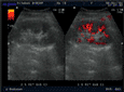
Ну конечно же, на представленных эхограммах - папиллярная опухоль мочевого пузыря.
Писать в заключении, что это полип - нельзя, это случай при котором руководствуются правилом - считаем опухоль злокачественной, пока не доказано обратное. Опухоль расположена над устьем левого мочеточника, нет обструкции устья и терминального отдела мочеточника (доказательство - наличие полноценного выброса мочи из устья) - что, в свою очередь, говорит против интралюминальной уретеральной опухоли.
Писать в заключении, что это полип - нельзя, это случай при котором руководствуются правилом - считаем опухоль злокачественной, пока не доказано обратное. Опухоль расположена над устьем левого мочеточника, нет обструкции устья и терминального отдела мочеточника (доказательство - наличие полноценного выброса мочи из устья) - что, в свою очередь, говорит против интралюминальной уретеральной опухоли.
-
KsV
- Администратор
- Сообщения: 2192
- Зарегистрирован: 22 фев 2009, 13:38
- Откуда: Беларусь-Lietuva
- Контактная информация:
Re: УЗИ мочевого пузыря
Именно ради "основополагающей заповеди врача - Не навреди" оно и существует. Так как невозможно в таких случаях на основании только ультразвуковых данных исключить карциному.Uzgraph писал(а):Кто является автором этого правила?KapustinSV писал(а): Писать в заключении, что это полип - нельзя, это случай при котором руководствуются правилом - считаем опухоль злокачественной, пока не доказано обратное.
И ещё один вопрос, если администратор разрешит его себе задать, а вам не кажется, что данным правилом нарушается одна из основополагающих заповедей врача - Не навреди. Незнание знанием не является! Как врач придерживающийся естественнонаучных принципов вы должны это знать. Поэтому в следующий раз, когда захотите безосновательно попугать пациента, лишний раз подумайте, мой вам совет.
Тактика одна - ТУР --> гистология.

Re: УЗИ мочевого пузыря
Дискуссия на эту тему не нова, статистики и исследование у разных авторов довольно широко варьируют. Если иcxодить из постулата: на навреди, то давайте порассуждаем так: можно ли на основании одного УЗИ (да и КТ так само) быть уверенным в доброкачественности или злокачественности экзофитного образования мочевого пузыря? Понятно что нельзя. Далее, какова должна быть тогда дальнейшая тактика? Опять же, опираясь на постулат "не навреди". Наблюдать? Гм, исходя из того, что рак это очень опасное заболевание, наблюдать как он растет переходя из одной стадии в другую, опасно для здоровья не только пациента, но иногда и самого "наблюдающего". Даже при такой жизнеутверждающей статистике, которую привёл коллега Uzograph. По тому же принципу, на фиг тогда мы делаем маммографию и заказываем биопсию при BIRADS 4? 
Ладно, что бы не быть голословным (только не обвиняйте в плагиате!), приведу выдержки и главы 21 Smith's General Urology 17th edition 2008. Книга и сам автор довольно авторитетны, и уже последние 25 лет являются настольным справочником для урологов в США и Европы. Так вот что там пишут:
Urothelial Carcinoma: Cancers of
the Bladder, Ureter, & Renal Pelvis
Badrinath R. Konety, MD, MBA, & Peter R. Carroll, MD
BLADDER CARCINOMAS
Incidence
Bladder cancer is the second most common cancer of the
genitourinary tract. It accounts for 7% of new cancer cases
in men and 2% of new cancer cases in women. The incidence
is higher in whites than in African Americans, and
there is a positive social class gradient for bladder cancer in
both sexes. The average age at diagnosis is 65 years. At that
time, approximately 75% of bladder cancers are localized
to the bladder; 25% have spread to regional lymph nodes
or distant sites.
Histopathology
Ninety-eight percent of all bladder cancers are epithelial
malignancies, with most being transitional cell carcinomas
(TCCs). !!!!!!!!!!!!!!!!!!!!!!!!!!!!!!!!!!!!!!!!!!!!!!!!!!!!!!!!! (Восклицательные знаки мои, перевести или и так понятно?)
A. NORMAL UROTHELIUM
The normal urothelium is composed of 3–7 layers of transitional
cell epithelium resting on a basement membrane
composed of extracellular matrix (collagen, adhesive glycoproteins,
glycosaminoglycans). The epithelial
cells vary in appearance: The basal cells are actively
proliferating cells resting on the basement membrane; the
luminal cells, perhaps the most important feature of normal
bladder epithelium, are larger umbrella-like cells that
are bound together by tight junctions. Beyond the basement
membrane is loose connective tissue, the lamina propria,
in which occasionally smooth-muscle fibers can be
identified. These fibers should be distinguished from
deeper, more extensive muscle elements defining the true
muscularis propria. The muscle wall of the bladder is composed
of muscle bundles coursing in multiple directions.
As these converge near the bladder neck, 3 layers can be
recognized: inner and outer longitudinally oriented layers
and a middle circularly oriented layer.
B. PAPILLOMA
The World Health Organization recognizes a papilloma
as a papillary tumor with a fine fibrovascular stalk supporting
an epithelial layer of transitional cells with normal
thickness and cytology (Epstein et al, 1998). Papillomas
are a rare benign condition usually occurring in
younger patients. Rare по английски-РЕДКО, .е. паппилома это редкая патология, в основном у молодых пациентов.

C. TRANSITIONAL CELL CARCINOMA
Approximately 90% of all bladder cancers are TCCs.
These tumors most commonly appear as papillary, exophytic
lesions; less commonly, they may
be sessile or ulcerated. Whereas the former group is usually
superficial in nature, sessile growths are often invasive.
Carcinoma in situ (CIS) is recognizable as flat, anaplastic
epithelium. The urothelium lacks the normal cellular
polarity, and cells contain large, irregular hyperchromatic
nuclei with prominent nucleoli.
D. NONTRANSITIONAL CELL CARCINOMAS
1. Adenocarcinoma—Adenocarcinomas account for
<2% of all bladder cancers. Primary adenocarcinomas of
the bladder may be preceded by cystitis and metaplasia.
Histologically, adenocarcinomas are mucus-secreting
and may have glandular, colloid, or signet-ring patterns.
Whereas primary adenocarcinomas often arise along the
floor of the bladder, adenocarcinomas arising from the urachus
occur at the dome. Both tumor types are often localized
at the time of diagnosis, but muscle invasion is usually
present. Five-year survival is usually <40%, despite aggressive
surgical management (Kramer et al, 1979; Abenoza,
Manivel, and Fraley, 1987; Bernstein et al, 1988).
2. Squamous cell carcinoma—Squamous cell carcinoma
accounts for between 5% and 10% of all bladder
cancers in the United States and is often associated here
with a history of chronic infection, vesical calculi, or
chronic catheter use. It may also be associated with bilharzial
infection owing to Schistosoma haematobium, because
squamous cell carcinoma accounts for approximately 60%
of all bladder cancers in Egypt, parts of Africa, and the
Middle East, where this infection is prevalent (El-Bolkainy
et al, 1981). These tumors are often nodular and invasive
at the time of diagnosis. Histologically they appear as
poorly differentiated neoplasms composed of polygonal
cells with characteristic intercellular bridges. Keratinizing
epithelium is present, although often in small amounts.
3. Undifferentiated carcinomas—Undifferentiated
bladder carcinomas, which are rare (accounting for <2%),
have no mature epithelial elements. Very undifferentiated
tumors with neuroendocrine features and small cell carcinomas
tend to be aggressive and present with metastases
(Quek et al, 2005; Choong et al, 2005).
4. Mixed carcinoma—Mixed carcinomas constitute 4–
6% of all bladder cancers and are composed of a combination
of transitional, glandular, squamous, or undifferentiated
patterns. The most common type comprises transitional
and squamous cell elements (Murphy, 1989). Most
mixed carcinomas are large and infiltrating at the time of
diagnosis.
E. RARE EPITHELIAL & NONEPITHELIAL CANCERS
Rare epithelial carcinomas identified in the bladder include
villous adenomas, carcinoid tumors, carcinosarcomas, and
melanomas. Rare nonepithelial cancers of the urinary bladder
include pheochromocytomas, lymphomas, choriocarcinomas,
and various mesenchymal tumors (hemangioma,
osteogenic sarcoma, and myosarcoma) (Murphy, 1989).
Cancers of the prostate, cervix, and rectum may involve
the bladder by direct extension. The most common
tumors metastatic to the bladder include (in order of incidence)
melanoma, lymphoma, stomach, breast, kidney,
lung and liver (Murphy, 1989; Goldstein, 1967, Franks,
1999).
D. IMAGING
Although bladder cancers may be detected by various
imaging techniques, their presence is confirmed by cystoscopy
and biopsy. Imaging is therefore used to evaluate the
are detected, to assess the depth of muscle wall infiltration
and the presence of regional or distant metastases. Intravenous
urography remains one of the most common imaging
tests for the evaluation of hematuria. However, intravenous
pyelography is increasingly being replaced by
computed tomography (CT) urography, which is more
accurate, for evaluation of the entire abdominal cavity,
renal parenchyma, and ureters in patients with hematuria
(Gray Sears et al, 2002). Bladder tumors may be recognized
as pedunculated, radiolucent filling defects projecting
into the lumen; nonpapillary, infiltrating
tumors may result in fixation or flattening of the
bladder wall. Hydronephrosis from ureteral obstruction is
usually associated with deeply infiltrating lesions and poor
outcome after treatment (Haleblian et al, 1998).
Superficial (Ta, Tis) bladder cancers staged with a
properly performed TUR and examination under anesthesia
do not require additional imaging of the bladder or pelvic
organs.
Т.е. автор сообщает что экзофитные, поверхностные опухоли достаточно диагностировать на основании ТУР, без дополнительной визуализации.

However, higher stage lesions are often understaged,
and the addition of imaging may be useful. Both
CT and magnetic resonance imaging (MRI)
have been used to characterize the extent of bladder wall
invasion and detect enlarged pelvic lymph nodes, with
overall staging accuracy ranging from 40% to 85% for CT
and from 50% to 90% for MRI (Fisher, Hricak, and Tanagho,
1985; Wood et al, 1988). Both techniques rely on
size criteria for the detection of lymphadenopathy: Lymph
nodes >1 cm are thought to be suggestive of metastases;
unfortunately, small-volume pelvic lymph node metastases
are often missed. Because invasive bladder cancers may
metastasize to the lung or bones, staging of advanced
lesions is completed with chest x-ray and radionuclide
bone scan. Bone scans can be avoided if the serum alkaline
phosphatase is normal (Berger, 1981).
E. CYSTOURETHROSCOPY & TUMOR RESECTION
The diagnosis and initial staging of bladder cancer is made
by cystoscopy and transurethral resection (TUR). Cystoscopy
can be done with either flexible or rigid instruments,
although the former is associated with less discomfort and
only requires local anesthesia. Superficial, low-grade tumors
usually appear as single or multiple papillary lesions. Higher
grade lesions are larger and sessile. CIS may appear as flat
areas of erythema and mucosal irregularity. Use of fluorescent
cystoscopy with blue light can enhance the ability to
detect lesions by as much as 20% (Jocham, 2005). In this
procedure, hematoporphyrin derivatives that accumulate
preferentially in cancer cells are instilled into the bladder
and fluorescence incited using a blue light. Cancer cells
with accumulated porphyrin such as 5-aminolevulenic acid
or hexaminolevulinate (HAL) are detected as glowing red
under the fluorescent light (Loidl, 2005).
Once a tumor is visualized or suspected, the patient is
scheduled for examination under anesthesia and TUR or
biopsy of the suspicious lesion. The objectives are tumor
diagnosis, assessment of the degree of bladder wall invasion
(staging), and complete excision of the low-stage lesions
amenable to such treatment.
Patients are placed in the lithotomy position. A careful
bimanual examination is performed. The presence of any
palpable mass and mobility of the bladder are noted, along
with any degree of fixation to contiguous structures. Cystoscopy
is repeated with one or more lenses (30° and 70°)
that permit complete visualization of the entire bladder
surface. A resectoscope is then placed into the bladder, and
visible tumors are removed by electrocautery. Suspicious
areas may be biopsied with cup biopsy forceps and the
areas may be cauterized with an electrode. Some clinicians
routinely perform random bladder biopsies of normalappearing
urothelium both close to and remote from the
tumor. The value of random bladder biopsies is controversial.
Detection of CIS on these biopsies can alter treatment
though more recent studies suggest that only 1.5% of lowrisk
and 3.5% of high-risk patients may have tumor
detected on such biopsies. (van der Meijden, 1999; May et
al, 2003). Findings of the random biopsy can alter treatment
in up to 7% of patients (May et al, 2003).
Кажется хватит; надеюсь что такой уважаемый источник достаточно внятно и убедительно докзывает, что только ТУР с биопсией является достоверным диагностичким приёмом, и пока по другому быть не может. Так что "не навреди", если есть образование мочевого пузыря, есть риск что рак, значит надо выполнить цистоскопию и взять биопсию.
С Уважением ко всем участникам Др. Мaрио.
N.B. если есть проблемы с переводом, спрашивайте, помогу.
Ладно, что бы не быть голословным (только не обвиняйте в плагиате!), приведу выдержки и главы 21 Smith's General Urology 17th edition 2008. Книга и сам автор довольно авторитетны, и уже последние 25 лет являются настольным справочником для урологов в США и Европы. Так вот что там пишут:
Urothelial Carcinoma: Cancers of
the Bladder, Ureter, & Renal Pelvis
Badrinath R. Konety, MD, MBA, & Peter R. Carroll, MD
BLADDER CARCINOMAS
Incidence
Bladder cancer is the second most common cancer of the
genitourinary tract. It accounts for 7% of new cancer cases
in men and 2% of new cancer cases in women. The incidence
is higher in whites than in African Americans, and
there is a positive social class gradient for bladder cancer in
both sexes. The average age at diagnosis is 65 years. At that
time, approximately 75% of bladder cancers are localized
to the bladder; 25% have spread to regional lymph nodes
or distant sites.
Histopathology
Ninety-eight percent of all bladder cancers are epithelial
malignancies, with most being transitional cell carcinomas
(TCCs). !!!!!!!!!!!!!!!!!!!!!!!!!!!!!!!!!!!!!!!!!!!!!!!!!!!!!!!!! (Восклицательные знаки мои, перевести или и так понятно?)
A. NORMAL UROTHELIUM
The normal urothelium is composed of 3–7 layers of transitional
cell epithelium resting on a basement membrane
composed of extracellular matrix (collagen, adhesive glycoproteins,
glycosaminoglycans). The epithelial
cells vary in appearance: The basal cells are actively
proliferating cells resting on the basement membrane; the
luminal cells, perhaps the most important feature of normal
bladder epithelium, are larger umbrella-like cells that
are bound together by tight junctions. Beyond the basement
membrane is loose connective tissue, the lamina propria,
in which occasionally smooth-muscle fibers can be
identified. These fibers should be distinguished from
deeper, more extensive muscle elements defining the true
muscularis propria. The muscle wall of the bladder is composed
of muscle bundles coursing in multiple directions.
As these converge near the bladder neck, 3 layers can be
recognized: inner and outer longitudinally oriented layers
and a middle circularly oriented layer.
B. PAPILLOMA
The World Health Organization recognizes a papilloma
as a papillary tumor with a fine fibrovascular stalk supporting
an epithelial layer of transitional cells with normal
thickness and cytology (Epstein et al, 1998). Papillomas
are a rare benign condition usually occurring in
younger patients. Rare по английски-РЕДКО, .е. паппилома это редкая патология, в основном у молодых пациентов.
C. TRANSITIONAL CELL CARCINOMA
Approximately 90% of all bladder cancers are TCCs.
These tumors most commonly appear as papillary, exophytic
lesions; less commonly, they may
be sessile or ulcerated. Whereas the former group is usually
superficial in nature, sessile growths are often invasive.
Carcinoma in situ (CIS) is recognizable as flat, anaplastic
epithelium. The urothelium lacks the normal cellular
polarity, and cells contain large, irregular hyperchromatic
nuclei with prominent nucleoli.
D. NONTRANSITIONAL CELL CARCINOMAS
1. Adenocarcinoma—Adenocarcinomas account for
<2% of all bladder cancers. Primary adenocarcinomas of
the bladder may be preceded by cystitis and metaplasia.
Histologically, adenocarcinomas are mucus-secreting
and may have glandular, colloid, or signet-ring patterns.
Whereas primary adenocarcinomas often arise along the
floor of the bladder, adenocarcinomas arising from the urachus
occur at the dome. Both tumor types are often localized
at the time of diagnosis, but muscle invasion is usually
present. Five-year survival is usually <40%, despite aggressive
surgical management (Kramer et al, 1979; Abenoza,
Manivel, and Fraley, 1987; Bernstein et al, 1988).
2. Squamous cell carcinoma—Squamous cell carcinoma
accounts for between 5% and 10% of all bladder
cancers in the United States and is often associated here
with a history of chronic infection, vesical calculi, or
chronic catheter use. It may also be associated with bilharzial
infection owing to Schistosoma haematobium, because
squamous cell carcinoma accounts for approximately 60%
of all bladder cancers in Egypt, parts of Africa, and the
Middle East, where this infection is prevalent (El-Bolkainy
et al, 1981). These tumors are often nodular and invasive
at the time of diagnosis. Histologically they appear as
poorly differentiated neoplasms composed of polygonal
cells with characteristic intercellular bridges. Keratinizing
epithelium is present, although often in small amounts.
3. Undifferentiated carcinomas—Undifferentiated
bladder carcinomas, which are rare (accounting for <2%),
have no mature epithelial elements. Very undifferentiated
tumors with neuroendocrine features and small cell carcinomas
tend to be aggressive and present with metastases
(Quek et al, 2005; Choong et al, 2005).
4. Mixed carcinoma—Mixed carcinomas constitute 4–
6% of all bladder cancers and are composed of a combination
of transitional, glandular, squamous, or undifferentiated
patterns. The most common type comprises transitional
and squamous cell elements (Murphy, 1989). Most
mixed carcinomas are large and infiltrating at the time of
diagnosis.
E. RARE EPITHELIAL & NONEPITHELIAL CANCERS
Rare epithelial carcinomas identified in the bladder include
villous adenomas, carcinoid tumors, carcinosarcomas, and
melanomas. Rare nonepithelial cancers of the urinary bladder
include pheochromocytomas, lymphomas, choriocarcinomas,
and various mesenchymal tumors (hemangioma,
osteogenic sarcoma, and myosarcoma) (Murphy, 1989).
Cancers of the prostate, cervix, and rectum may involve
the bladder by direct extension. The most common
tumors metastatic to the bladder include (in order of incidence)
melanoma, lymphoma, stomach, breast, kidney,
lung and liver (Murphy, 1989; Goldstein, 1967, Franks,
1999).
D. IMAGING
Although bladder cancers may be detected by various
imaging techniques, their presence is confirmed by cystoscopy
and biopsy. Imaging is therefore used to evaluate the
are detected, to assess the depth of muscle wall infiltration
and the presence of regional or distant metastases. Intravenous
urography remains one of the most common imaging
tests for the evaluation of hematuria. However, intravenous
pyelography is increasingly being replaced by
computed tomography (CT) urography, which is more
accurate, for evaluation of the entire abdominal cavity,
renal parenchyma, and ureters in patients with hematuria
(Gray Sears et al, 2002). Bladder tumors may be recognized
as pedunculated, radiolucent filling defects projecting
into the lumen; nonpapillary, infiltrating
tumors may result in fixation or flattening of the
bladder wall. Hydronephrosis from ureteral obstruction is
usually associated with deeply infiltrating lesions and poor
outcome after treatment (Haleblian et al, 1998).
Superficial (Ta, Tis) bladder cancers staged with a
properly performed TUR and examination under anesthesia
do not require additional imaging of the bladder or pelvic
organs.
Т.е. автор сообщает что экзофитные, поверхностные опухоли достаточно диагностировать на основании ТУР, без дополнительной визуализации.
However, higher stage lesions are often understaged,
and the addition of imaging may be useful. Both
CT and magnetic resonance imaging (MRI)
have been used to characterize the extent of bladder wall
invasion and detect enlarged pelvic lymph nodes, with
overall staging accuracy ranging from 40% to 85% for CT
and from 50% to 90% for MRI (Fisher, Hricak, and Tanagho,
1985; Wood et al, 1988). Both techniques rely on
size criteria for the detection of lymphadenopathy: Lymph
nodes >1 cm are thought to be suggestive of metastases;
unfortunately, small-volume pelvic lymph node metastases
are often missed. Because invasive bladder cancers may
metastasize to the lung or bones, staging of advanced
lesions is completed with chest x-ray and radionuclide
bone scan. Bone scans can be avoided if the serum alkaline
phosphatase is normal (Berger, 1981).
E. CYSTOURETHROSCOPY & TUMOR RESECTION
The diagnosis and initial staging of bladder cancer is made
by cystoscopy and transurethral resection (TUR). Cystoscopy
can be done with either flexible or rigid instruments,
although the former is associated with less discomfort and
only requires local anesthesia. Superficial, low-grade tumors
usually appear as single or multiple papillary lesions. Higher
grade lesions are larger and sessile. CIS may appear as flat
areas of erythema and mucosal irregularity. Use of fluorescent
cystoscopy with blue light can enhance the ability to
detect lesions by as much as 20% (Jocham, 2005). In this
procedure, hematoporphyrin derivatives that accumulate
preferentially in cancer cells are instilled into the bladder
and fluorescence incited using a blue light. Cancer cells
with accumulated porphyrin such as 5-aminolevulenic acid
or hexaminolevulinate (HAL) are detected as glowing red
under the fluorescent light (Loidl, 2005).
Once a tumor is visualized or suspected, the patient is
scheduled for examination under anesthesia and TUR or
biopsy of the suspicious lesion. The objectives are tumor
diagnosis, assessment of the degree of bladder wall invasion
(staging), and complete excision of the low-stage lesions
amenable to such treatment.
Patients are placed in the lithotomy position. A careful
bimanual examination is performed. The presence of any
palpable mass and mobility of the bladder are noted, along
with any degree of fixation to contiguous structures. Cystoscopy
is repeated with one or more lenses (30° and 70°)
that permit complete visualization of the entire bladder
surface. A resectoscope is then placed into the bladder, and
visible tumors are removed by electrocautery. Suspicious
areas may be biopsied with cup biopsy forceps and the
areas may be cauterized with an electrode. Some clinicians
routinely perform random bladder biopsies of normalappearing
urothelium both close to and remote from the
tumor. The value of random bladder biopsies is controversial.
Detection of CIS on these biopsies can alter treatment
though more recent studies suggest that only 1.5% of lowrisk
and 3.5% of high-risk patients may have tumor
detected on such biopsies. (van der Meijden, 1999; May et
al, 2003). Findings of the random biopsy can alter treatment
in up to 7% of patients (May et al, 2003).
Кажется хватит; надеюсь что такой уважаемый источник достаточно внятно и убедительно докзывает, что только ТУР с биопсией является достоверным диагностичким приёмом, и пока по другому быть не может. Так что "не навреди", если есть образование мочевого пузыря, есть риск что рак, значит надо выполнить цистоскопию и взять биопсию.
С Уважением ко всем участникам Др. Мaрио.
N.B. если есть проблемы с переводом, спрашивайте, помогу.
-
KsV
- Администратор
- Сообщения: 2192
- Зарегистрирован: 22 фев 2009, 13:38
- Откуда: Беларусь-Lietuva
- Контактная информация:
Re: УЗИ мочевого пузыря
Так все-таки, наблюдаем?Uzgraph писал(а):KapustinSV писал(а):...
Таким, образом, TCC необходимо наблюдать?
Скажите, а мы лечим ТСС или человека?
-
KsV
- Администратор
- Сообщения: 2192
- Зарегистрирован: 22 фев 2009, 13:38
- Откуда: Беларусь-Lietuva
- Контактная информация:
Re: УЗИ мочевого пузыря
Так, все таки, не понятно, Uzgraph, как, по Вашему, нужно поступать с такими пациентами? Проясните свою точку зрения, пожалуйста.
Кто сейчас на конференции
Сейчас этот форум просматривают: нет зарегистрированных пользователей и 1 гость